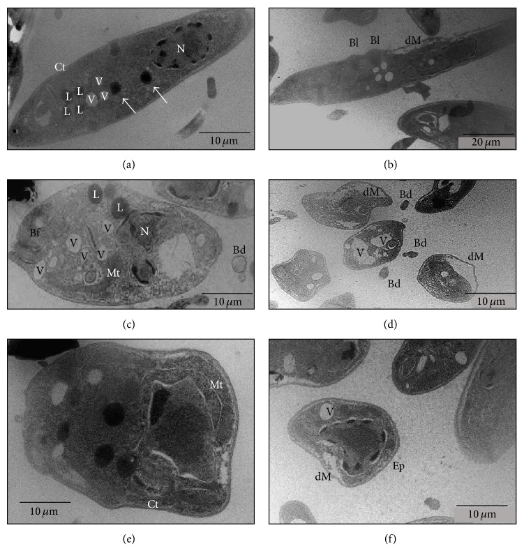
Figure 3.
Transmission electron micrographs promastigote forms of Leishmania infantum chagasi. (a) Control group; (b), (c), (d), (e), and (f) treated groups (AU) at a concentration of 25 μg/mL for 72 hours. Vacuoles (V), acidocalcisomes (arrows), nucleus (N), lipid (L), kinetoplast (Ct), flagellar pocket (Bf), mitochondrial swelling (Mt), detachment of the plasma membrane (dM), blebs (Bl), detached blebs (Bd), and electron-lucent space (Ep). Bars 10 μm (a, c, d, e, and f), Bars 20 μm (b).

