
Fig 1. Stx2-containing blood cell-derived microvesicles detected in the renal cortex of a HUS patient.
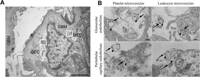
(A) Ultramorphology showing an overview from the renal cortex of a patient with HUS (patient 14) including a glomerular capillary with two boxes magnified in (B). Scale bar: 2 μm. P: podocyte, GEC: glomerular endothelial cell, GBM: glomerular basement membrane, L: leukocyte. (B) Renal cortex of a the same HUS patient labeled with anti-Stx2 (5 nm, arrowhead), anti-CD42b (10 nm, arrow) to detect platelet-derived microvesicles or anti-CD45 (10 nm, arrow) to detect leukocyte-derived microvesicles. Microvesicles were defined by their size (shed microvesicles: ≤ 1μm, apoptotic bodies: 1–5 μm) and their cellular origin based on the presence of platelet- or leukocyte-markers. Stx2-containing microvesicles were detected in glomerular- and peritubular capillary endothelium. The two upper panels are magnifications of the two boxes depicted in (A). Scale bar: 100 nm.