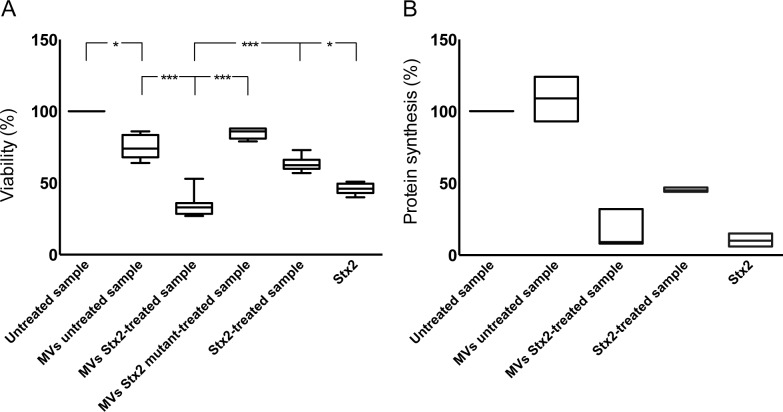
Fig 5. Microvesicles containing Stx2 affected the viability and inhibited protein synthesis in CiGEnC.
The effect of Stx2-containing microvesicles on cell viability was examined by use of a crystal violet assay (A). Data are expressed as percentage of control cell viability defined as 100%. Representative data from five experiments are depicted as the median of triplicate wells. *** Denotes P value <0.001 and * P<0.05. (B) Protein synthesis was measured as the incorporation of [35S]-methionine into total protein. Data are expressed as percent and 100% represents the untreated cells incubated under the same conditions but without microvesicles. All experiments were done in duplicate and the experiment was repeated three times. MVs untreated sample: whole blood was exposed to PBS for 1 hr, microvesicles (MVs) were isolated and incubated with CiGEnC for 36 hr; MVs Stx2-treated sample: isolated from whole blood treated with Stx2 (200 ng/mL diluted in PBS) for 36h; MVs Stx2 mutant-treated sample: isolated from whole blood treated with Stx2 mutated in the catalytic A subunit for 36h: Stx2-treated sample: purified Stx2 was exposed to washing steps similar to microvesicles before exposure to the cells; Stx2: cells were exposed to pure Stx2 (200 ng/mL) without washing steps.

