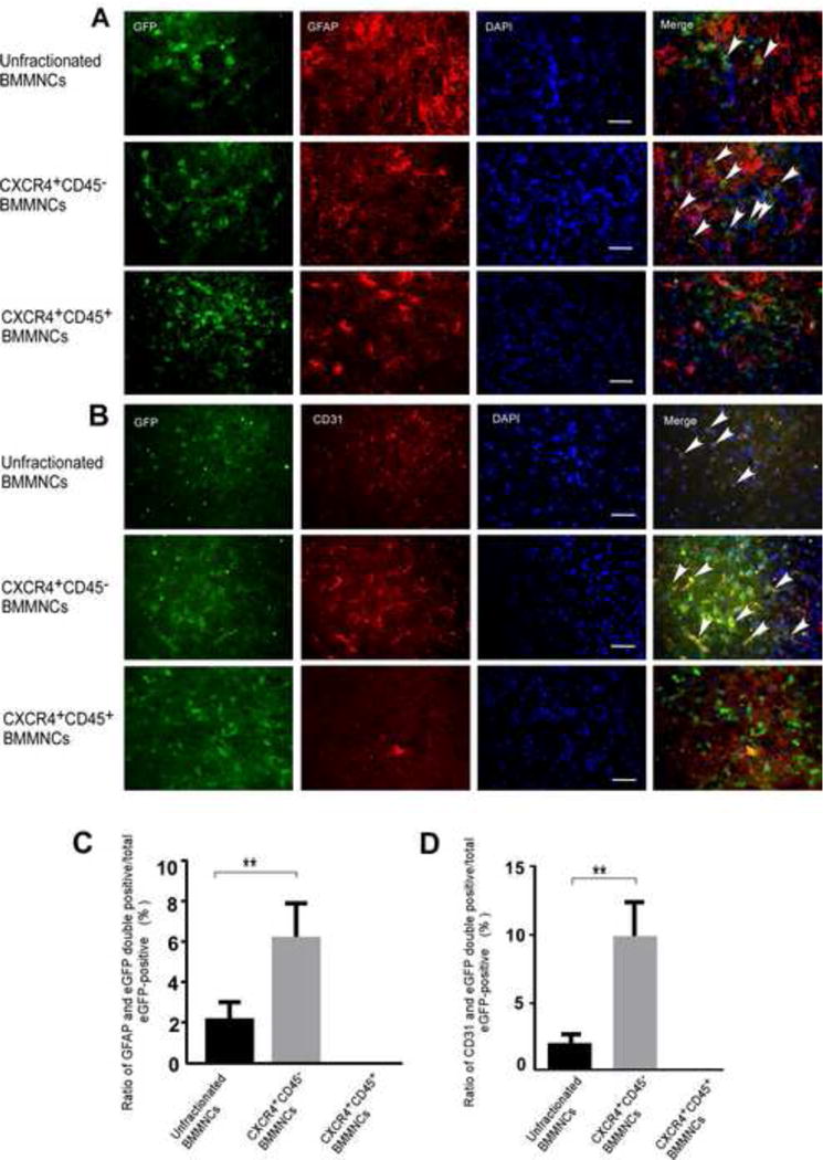
Figure 4. CXCR4+CD45− BMMNCs are more plastic than unfractionated BMMNCs in vivo.

Immunofluorescence staining from unfractionated BMMNC-treated mice (upper panel) and CXCR4+CD45− BMMNC-treated mice (middle panel) show that eGFP colocalizes with astrocyte marker GFAP (A) and endothelial cell marker CD31 (B). In ischemic brain tissue from CXCR4+CD45+ BMMNC-treated tMCAO mice (lower panel of A and B) eGFP did not colocalize with either cell-specific marker; scale bar = 50 μm. Arrows indicate cells with colocalization of eGFP and the cell-specific marker. (C and D) Quantification of the ratio of double-labeled cells that express GFAP (C) or CD31 (D) to total number of eGFP+ cells. n=6 mice/group; **p<0.01 vs. unfractionated BMMNCs; one-way ANOVA followed by Tamhane’s T2 test.
