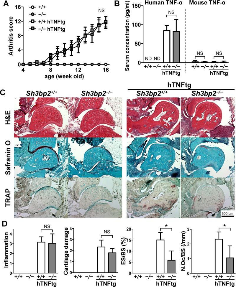
Figure 1.
Decreased osteoclast formation and bone erosion in SH3BP2-deficient human TNF-α-transgenic mice. Sh3bp2+/+ and Sh3bp2−/− mice were crossed with human TNF-α transgenic (hTNFtg) mice. Joint inflammation was monitored until the age of 16 weeks. Serum and hindlimbs were collected and subjected to ELISA and histological analysis, respectively. A, Changes in clinically assessed joint inflammation scores in the Sh3bp2+/+ (n = 9), Sh3bp2−/− (n = 7), Sh3bp2+/+/hTNFtg (n = 7), and Sh3bp2−/−/hTNFtg (n = 9) male mice. B, Serum concentrations of human and mouse TNF-α. C, Representative staining images of the ankle joint tissues. Ankle joint sections were stained with hematoxylin and eosin (H&E), Safranin O, and tartrate-resistant acid phosphatase (TRAP). Original magnification: 40X. D, Histological scores of inflammation and cartilage damage and histomorphometric analysis of talar bones. Bone erosion on the surface of the talus was traced, and attached osteoclasts were counted. Eroded surface per bone surface (ES/BS) and number of osteoclast per bone surface (N.Oc/BS) of the talus were determined. Values are presented as the mean ± SEM. +/+ = Sh3bp2+/+; −/− = Sh3bp2−/−. * = P < 0.05; NS = not significant; ND = not detectable.

