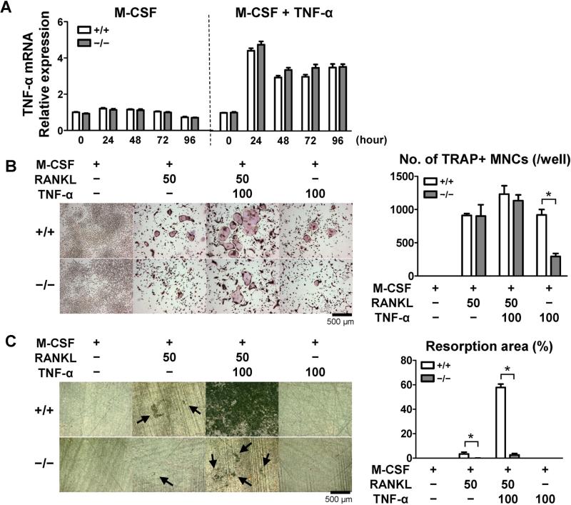
Figure 3.
Impaired osteoclast differentiation and bone-resorbing function in Sh3bp2−/− bone marrow-derived macrophages. Primary bone marrow cells were isolated and cultured as described in Methods section. A, TNF-α mRNA expression. Bone marrow-derived macrophages (BMMs) were stimulated with TNF-α (100 ng/ml) in the presence of M-CSF (25 ng/ml). TNF-α mRNA expression levels relative to Hprt were calculated and normalized to the expression level of Sh3bp2+/+ BMMs at 0 hour. B, Representative TRAP staining images and number of TRAP-positive multinucleated cells (TRAP+ MNCs). BMMs were stimulated with RANKL (50 ng/ml) and/or TNF-α (100 ng/ml) in the presence of M-CSF (25 ng/ml) for 4 days. Original magnification: 40X. C, Representative images and quantification of resorption area on dentine. BMMs were stimulated with RANKL (50 ng/ml) and/or TNF-α (100 ng/ml) in the presence of M-CSF (25 ng/ml) for 14 days. After removal of the cells, resorption areas were visualized by toluidine blue. Original magnification: 50X. Percentages of the resorption areas relative to total surface area were quantified.

