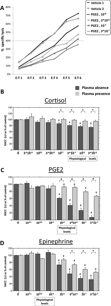
Fig. 1.
The effects of different hormones on NKCC against K562 target cells, in the presence of plasma and in its absence. (A) A representative E:T cytotoxicity curves in a single plate, including two replicates of control condition and 4 concentrations of PGE2, in the absence of plasma. Each curve is expressed as a single value (LU45, as explained in details in the Method section), and all results are presented as % of control levels of this index (1B–D). (B) Cortisol dose-dependently suppressed NKCC, but only in the absence of plasma. (C) PGE2 and (D) epinephrine dose-dependently suppressed NKCC, but this suppression was markedly more profound in the absence of plasma. ⍰ Indicates a significant difference between the presence and absence of plasma. # Indicates a significant reduction in NKCC compared to the respective control (Vehicle) levels. The concentrations of epinephrine and PGE2 spanned beyond systemic physiological levels, as these hormones, but not cortisol, also interact locally with NK cells at higher concentrations. Data presented as mean + SEM.

