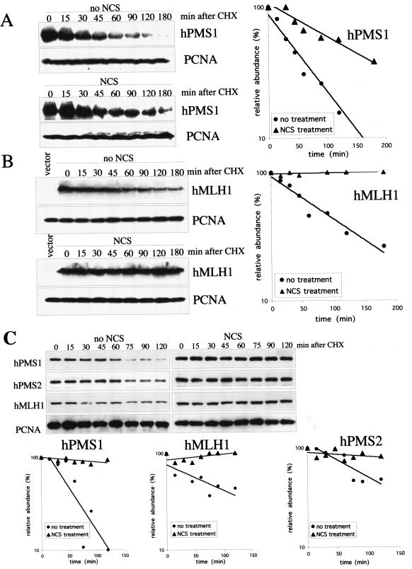
FIG. 4.
The half-lives of hMLH1, hPMS1, and hPMS2 are prolonged by NCS treatment. (A) HEK293T cells were transfected with pcDNA3- HA-hPMS1. Two days later, cells were either left untreated or treated with NCS (300 ng/ml) for 3 h followed by treatment with cycloheximide (CHX) at 20 μg/ml for the time periods as indicated. hPMS1 protein was detected by Western blot analysis using an antibody specific to hPMS1 (left panel) and quantified by densitometry (right panel). The relative abundance of hPMS1 following CHX treatment relative to that seen with no CHX treatment control was graphed (right panel). The PCNA immunoblot serves as a protein loading control. (B) HEK293T cells were transfected with pHcRed-hMLH1. The half-life of hMLH1 protein following CHX treatment was measured as described above. hMLH1 protein was detected by Western blot analysis with an hMLH1-specific antibody (left panels) and quantified by densitometry (right panel). (C) Growing HEK293 cells were either left untreated or treated with NCS (300 ng/ml) for 3 h followed by treatment with CHX (20 μg/ml) for the time periods as indicated. The endogenous hPMS1, hPMS2, and hMLH1 were detected by Western blot analysis (upper panels). The signals were quantified by densitometry, and the relative abundance of each protein following CHX treatment relative to that seen with the 0-min control was plotted (lower panels).

