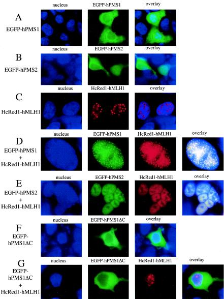
FIG. 6.
hMLH1 mediates nuclear localization of hPMS1 and hPMS2. HEK293 cells were transfected with expression plasmids as indicated on the left side of the figure. Cells were fixed, and nuclei were stained with Hoechst 33258. (A) When expressed alone, EGFP-hPMS1 was exclusively localized to the cytoplasm in 196 (49%) cells, distributed within the whole cells in 84 (21%) cells, and localized to the nucleus in 120 (30%) cells out of 400 transfected cells examined. (B) HEK293 cells were transfected with pEGFP-hPMS2. The distribution pattern showed that hPMS2 was located throughout the cells in 45%, was exclusively cytoplasmic in 23%, and was nuclear in 32% out of 400 GFP+ cells examined. (C) HcRed-hMLH1 was localized in the nucleus and formed discrete punctuates. The images reflect 396 (99%) of 400 transfected cell examined. Four cells exhibited diffuse nuclear distribution without the formation of foci. (D) Upon coexpression with hMLH1, EGFP-hPMS1 was translocated to nucleus and colocalized with HcRed1-hMLH1 in the nuclear foci in 400 (100%) out of 400 transfected cells examined. A total of 50% of cells contained numerous foci distributed throughout the nucleus as shown. The rest of the population exhibited a few nuclear foci (fewer than 20) in each nucleus. (E) HEK293 cells were cotransfected with pEGFP-hPMS2 and HcRed1-hMLH1. hPMS2 was exclusively localized to nucleus when it was coexpressed with HcRed1-hMLH1 in 400 (100%) out of 400 transfected cells examined. The images are representative of 99% of transfected cells. Only 1% of the cells contained more than 10 foci in the nucleus. (F) EGFP-hPMS1ΔC was exclusively localized in the cytoplasm. The images reflect 99% of 400 transfected cells examined. A total of 1% of the cells exhibited a whole-cell distribution pattern. (G) When coexpressed with hMLH1, EGFP-hPMS1ΔC remained in the cytoplasm. The images are representative of 99% of 400 transfected cells examined.

