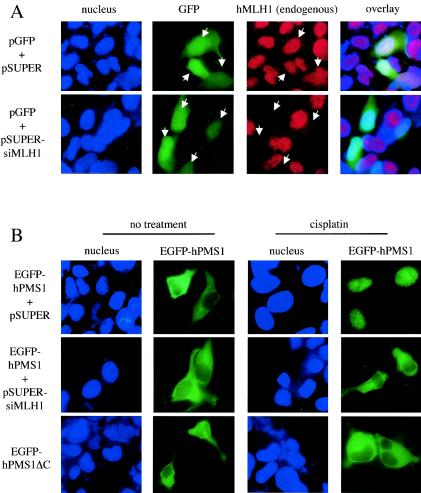
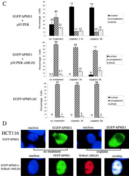
FIG. 7.
hMLH1 is required for nuclear localization of hPMS1 during DNA damage. (A) HEK293 cells were transfected with pGFP and either pSUPER (upper panels) or pSUPER-siMLH1 (lower panels). The cells were fixed, and hMLH1 was immunostained with its specific antibody followed by Texas Red X-conjugated anti-mouse secondary antibody. Nuclei were stained with Hoechst 33258. The transfected cells (arrows) were identified by the expression of GFP. Transfection of pSUPER empty vector did not alter the level of hMLH1 expression, but transfection of pSUPER-siMLH1 knocked down the expression of endogenous hMLH1. (B) HEK293 cells transfected with pEGFP-hPMS1 and either pSUPER (upper panels) or pSUPER-siMLH1 (middle panels) or transfected with pEGFP-hPMS1ΔC (lower panels) were treated with cisplatin (50 μM) for 3 h. Cells were fixed, and nuclei were stained with Hoechst 33258. Most EGFP-hPMS1 translocated from cytoplasm to nucleus and formed a nuclear punctate pattern upon cisplatin treatment (upper panels). MLH1 siRNA prevented the cisplatin-induced relocalization of EGFP-hPMS1 (middle panels). The hPMS1 mutant (hPMS1ΔC), lacking an hMLH1-interacting domain, remained in the cytoplasm following cisplatin treatment (lower panels). (C) GFP-positive cells of each sample in the experiments described for panel B were scored for the localization of EGFP-hPMS1 or EGFP-hPMS1ΔC, and the percentages of cells displaying exclusively nuclear, exclusively cytoplasmic, or both nuclear and cytoplasmic (whole) staining were determined. At least 300 GFP-positive cells were scored for each experiment. The data shown represent the means ± standard errors of three independent experiments. (D) HCT116 cells were transfected with the expression plasmids as indicated on the left. The cells were either left untreated or treated with cisplatin (50 μM) for 6 h. The cells were then fixed, and nuclei were stained with Hoechst 33258. EGFP-hPMS1 remained in the cytoplasm of hMLH1-deficient cells following cisplatin treatment but was relocalized to nucleus in hMLH1-reconstituted HCT116 cells. The images reflect 100% (upper panels) or 90% (lower panels) of 200 transfected cells examined. In hMLH1-reconstituted HCT116 cells, the distribution of hPMS1 was as follows: 90% exclusively nuclear, 4% exclusively cytoplasmic, and 6% in the whole cell.


