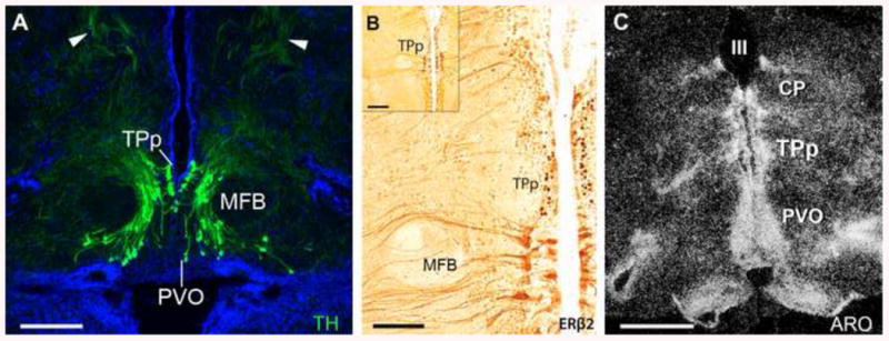
Fig. 11.

The dopaminergic periventricular posterior tuberculum (TPp) is an estrogen target in the midshipman brain. (A) Large, pear-shaped tyrosine hydroxylase immunoreactive (TH-ir; green) neurons, known to be dopaminergic, are found both in a true periventricular position just dorsal and lateral to the paraventricular organ (PVO), wrapping around the medial forebrain bundle (MFB) in a ventrolateral continuum. Arrowheads indicate thick dorsal projections that turn to descend through the brainstem. Blue is DAPI nuclear stain. For more details see [125]. (B) Robust ERβ2-ir (dark brown) in cells of the TPp. ERβ2-ir cells are found just medial to the MFB, similar to location of TH-ir neurons in A. Adapted from [49]. (C) High expression levels of aromatase mRNA (white grains) in the TPp and along the third ventricle (III) as visualized by dark-field in situ hybridization. CP, central posterior nucleus (auditory thalamus). Adapted from [50]. Scale bar = 250 μm in A, 100 μm in B, 200 μm in B inset, 500 μm in C.
