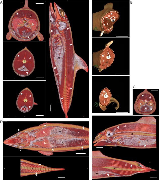
Fig 5. Distribution of brown adipose tissue in cetacean blubber.
(A) Whole body CT of Pacific white-side dolphin (SNH11012). Thoracic region (left upper panel), Abdominal region (left middle panel), Tail region (left lower panel). Lateral view of the body (right panel). (B) Whole body CT of bottlenose dolphin (UMUT-14006). The animal was scanned after necropsy. Thoracic region (upper panel), Abdominal region (middle panel), Gluteal region (lower panel). (C) Whole body CT of Dall's porpoise (SNH11011). Thoracic region (upper panel). Lateral view of the body (lower panel). (D) CT of Harbour porpoise (SNH10020). Horizontal section of the body. Arrows: thin layer of high-density/connective tissues in lower blubber layer. a, vertebrae; b, lung; c, heart; d, skeletal muscle; e, intestine; f, melon; g, brain; h, liver; i, thoracic cavity; j, abdominal cavity; k, eye. Scale bar = 10 cm.

