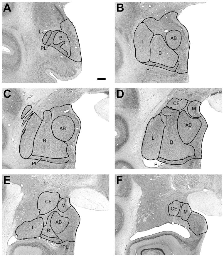
Figure 2.
Low-magnification photomicrographs of representative coronal sections through the monkey amygdala illustrating the locations of the main nuclei. L, lateral; B, basal; PL, paralaminar; AB, accessory basal; CE, central; M, medial. Nonlabeled areas represent remaining nuclei of the amygdala; see list in Table 1. Scale bar = 1 mm in A (applies to all panels).

