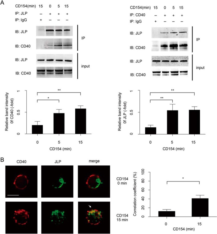
FIGURE 3.
JLP interacts with CD40 in B lymphocytes. A, cell extracts of splenic B lymphocytes from wild-type mice treated with rCD154 at the indicated time points were first incubated overnight with specific primary antibodies and then analyzed with the indicated antibody by co-immunoprecipitation. Input was 10% of total cell extracts, and immunoglobulin G was used as a negative control. Data are representative of three independent experiments. The indicated band intensity of immunoprecipitation (IP) was normalized to the relevant band intensity of the input. Means ± S.D. were calculated from three independent experiments. Error bars indicate S.D. *, p < 0.05; **, p < 0.01. IB, immunoblot. B, fluorescence microscopy of B lymphocytes from wild-type mice. Cells were untreated (0 min) or were treated with rCD154 for 15 min and then labeled with the indicated primary antibody followed by a fluorescence-conjugated secondary antibody. The white arrow indicates areas of overlap. Scale bar = 5 μm. Experiments were performed in triplicate. The co-localization correlation coefficients of CD40 and JLP were collected from three separate experiments and statistically analyzed using UltraVIEW software. *, p < 0.05 (Student's t test).

