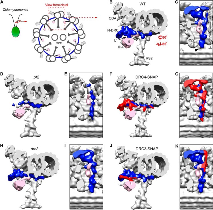
FIGURE 2.
SNAP-tagged DRC4 and DRC3 rescue the structural defects in pf2 and drc3. A, schematic of a Chlamydomonas axoneme in cross-sectional view, seen from the flagellar tip. ODA, IDA, and the N-DRC connect neighboring microtubule doublets, whereas the RS connect to the central pair complex (CPC). B–K, isosurface renderings of the three-dimensional structures of the 96-nm axonemal repeats in WT (B and C), pf2 (D and E), DRC4-SNAP (C-terminally SNAP-tagged DRC4, F and G), drc3 (H and I), and DRC3-SNAP (C-terminally SNAP-tagged DRC3, J and K) after cryo-ET and subtomogram averaging; cross-sectional views from distal (B, D, F, H, and J) and bottom views (C, E, G, I, and K) of the N-DRC (blue or red/blue). Note that the N-DRC defects are more severe in pf2 (D and E) than drc3 (H and I), but both the C-terminally SNAP-tagged DRC4 and DRC3 rescue strains, respectively, show recovery (colored red) of the N-DRC to WT morphology in DRC4-SNAP (F and G) and DRC3-SNAP (J and K). The hole in the A-B inner junction is indicated by an arrowhead (C). Other labels used are as follows: At, A-tubule; Bt, B-tubule; IDA e/g, inner dynein arms e and g; N-DRC, nexin-dynein regulatory complex; ODA, outer dynein arms; RS2, radial spoke 2; L1, L1 projection.

