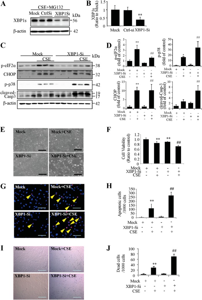
FIGURE 6.
Knockdown of XBP1 exacerbated CSE-induced cell apoptosis. ARPE-19 cells were transfected with XBP1 siRNA to knockdown XBP1 expression and then treated with CSE for 24 h. A, to determine the XBP1 siRNA knockdown efficiency, 10 μm of MG132, a proteasome inhibitor, was added to culture medium during the last 4 h of the CSE treatment. Western blotting shows down-regulation of XBP1s after XBP1 siRNA. B, quantification of XBP1 siRNA efficiency by densitometry. C, protein level of p-eIF2α, CHOP, p-p38 and cleaved-caspase-3 were determined by Western blotting. D, densitometry analysis of Western blots for p-eIF2α, CHOP, p-p38, and cleaved-caspase-3 (normalized with β-actin). E, representative images of siRNA-transfected ARPE-19 cells after CSE treatment for 24 h. Scale Bar: 200 μm. F, cell viability was quantified by MTT assay. G, apoptosis examined by TUNEL assay. Scale Bar: 100 μm. H, quantification of apoptotic cells number (TUNEL-positive cells) from G). I, cell death was detected using in situ Trypan Blue staining. Scale Bar: 200 μm. J, quantification of dead cells (Trypan Blue-stained cells). All data were expressed as mean ± S.D., from three independent experiments. *, p < 0.05; **, p < 0.01 versus mock; #, p < 0.05; ##, p < 0.01, versus mock + CSE.

