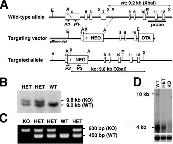
FIG. 2.
Targeted disruption of the TACC2 gene. (A) Schematic structure of and targeting strategy for the murine TACC2 locus. Empty boxes represent exons 4 to 12 of the murine TACC2 gene. The locations of the 5′ external probe and PCR primers for genotyping are indicated. Restriction enzyme sites are as follows: E, EcoRI; A, AflII; X, XbaI. wt, wild type; NEO, neomycin resistance cassette; DTA, diphtheria toxin A cassette; ko, knockout. Confirmation of successful targeting of ES cells was obtained by Southern blot analysis (B) and PCR screening of F1 mice for the presence of the disrupted allele (C). WT, wild type; KO, knockout; HET, heterozygote. (D) Northern blot analysis of kidney tissue with a TACC2-specific probe shows no hybridization signal in knockout tissue. Note that disruption of the TACC2 gene led to the complete absence of both the 4- and 10-kb transcripts, therefore generating a null mutation for all transcript variants. Hybridization for actin (lower panel) was used to control RNA loading. The positions of transcript sizes are indicated.

