
FIGURE 5.
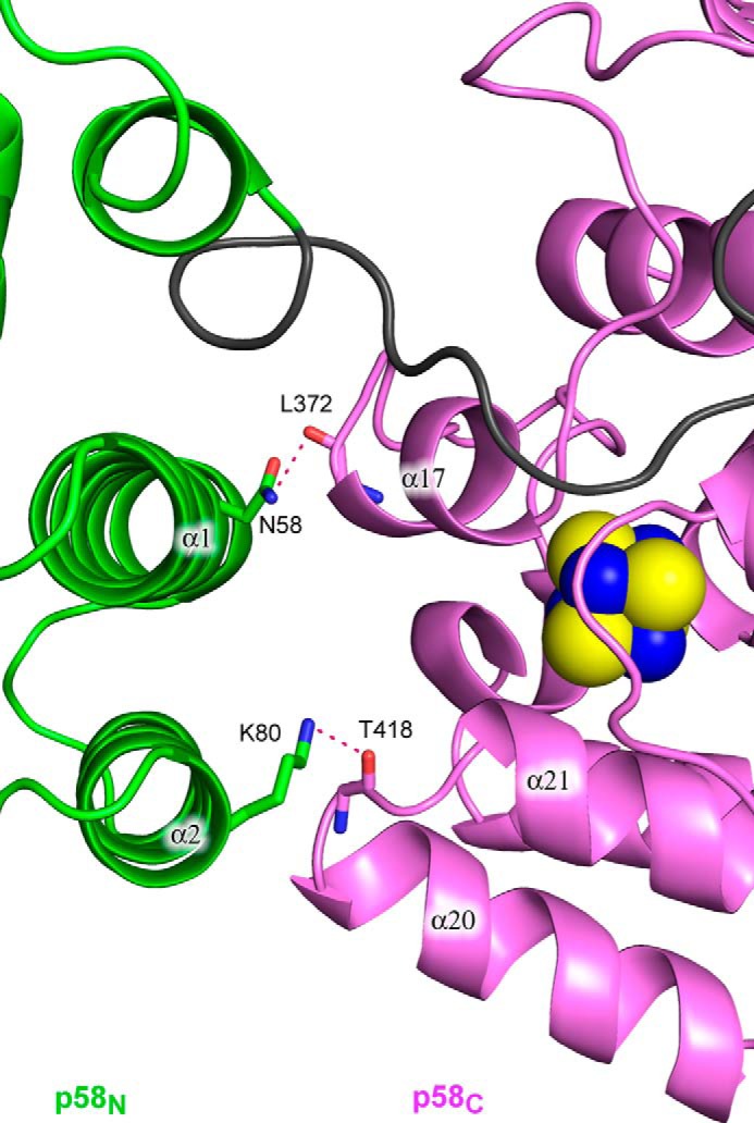
Close-up view of the p58N-p58C interaction interface. The protein is represented as a schematic and colored according to Fig. 1B. Side or main chains making the hydrogen bonds between p58N and p58C are shown as sticks. Hydrogen bonds are drawn with red dashed lines.