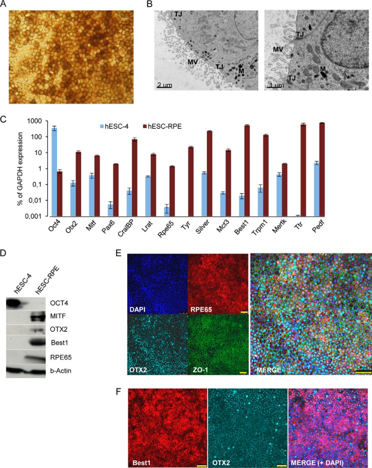
FIGURE 1.
Characterization of hESC-derived RPE cells. Pigmented clusters of polygonal cells were selected and reseeded onto Matrigel-coated plates. After 6 weeks of proliferation into high-density cultures, the cells reacquired the morphology and pigmentation of typical RPE cells (A). B, transmission electron microscopy of hESC-derived RPE cells showing typical RPE characteristics: apical microvilli (MV), melanin granules (M), and tight junctions (TJ). Scale bar = 2 μm (left panel) and 1 μm (right panel). C, qRT-PCR analysis of human ES cells and RPE-specific gene expression. Relative expression is shown as percentage of GAPDH expression. Data are presented as mean ± S.E. of three independent experiments performed in triplicate. D, Western blot analysis on cell lysates from human ES cells and hESC-RPE cells with antibodies against OCT4, MITF, OTX2, BEST1, RPE65, and β-Actin. Immunostaining shows coexpression of RPE65, OTX2, and ZO-1 (E) and BEST1 and OTX2 (F). DAPI was used to counterstain the nuclei (blue). Scale bars = 50 μm (E and F).

