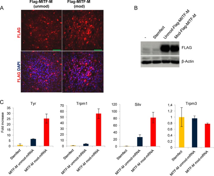
FIGURE 6.
Nuclear localization and functional expression of FLAG-MITF mRNA transfected into hESC-RPE cells. A, immunofluorescence staining of confluent hESC-RPE cells transfected for 16 h with unmod and mod mRNA encoding FLAG-MITF-M. DAPI (blue) was used to stain the nucleus. Scale bars = 100 μm. B, Western blots of early confluent cells 16 h post-transfection. C, qRT-PCR analysis of a subset of RPE-specific genes, including reported MITF-target genes (TYR, SILV, and TRPM1). Data are from one representative experiment of three independent experiments. Error bars represent mean ± S.D.

