
FIGURE 5.
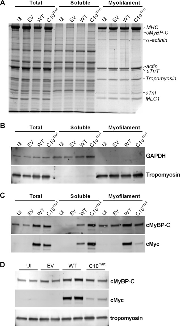
Determination of localization of AV-derived cMyBP-C with subcellular fractionation. Cultured cardiomyocytes were homogenized in buffer containing 1% Triton X-100 and centrifuged. Supernatant was named soluble, whereas the pellet was named myofilament fraction. A, Coomassie-stained SDS-polyacrylamide gel of total, soluble, and myofilament fractions. B, high fractionation efficiency was achieved as shown by using markers for cytosol (GAPDH) and myofilament (α-TPM). C, dual color immunoblots incubated with antibodies against cMyBP-C (endogenous + virus-derived cMyBP-C) and cMyc (virus-derived cMyBP-C). No cMyBP-C was detected in soluble fraction of uninfected (UI) or empty vector (EV) fractions, whereas it was detected in cMyBP-CWT and, especially, in cMyBP-CC10mut. cMyc signal shows the lower incorporation of cMyBP-CC10mut into the sarcomere. D, immunoblots of myofilament fractions of UI, EV, cMyBP-CWT, and cMyBP-CC10mut cells showed that cMyBP-CC10mut incorporated into the sarcomere at a lower level than cMyBP-CWT.