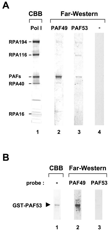
FIG. 4.
Far-Western analysis of PAF49 and PAF53. (A) Purified Pol IB was blotted onto a nitrocellulose membrane and then incubated with 35S-labeled PAF49 (lane 2) or PAF53 (lane 3) prepared by in vitro coupled transcription-translation reaction. A control labeling reaction with an empty plasmid template was used as control probe (lane 4). Same Pol I sample was also loaded onto the SDS-polyacrylamide gel, and individual subunits of Pol I were stained by Coomassie brilliant blue (lane 1). The positions of formerly identified subunits and PAFs are indicated at the left. (B) The far-Western blotting experiment was performed with purified GST-PAF53 and 35S-labeled PAF49 (lane 2) or PAF53 (lane 3). The purity of GST-PAF53 was indicated by Coomassie staining (lane 1).

