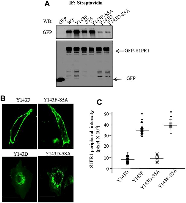
Fig. 4.
Y143-phosphorylation-dependent regulation of S1PR1 localization functions independently of serine phosphorylation of S1PR1. (A) CHO cells transiently transfected with the indicated GFP-tagged constructs were biotinylated. Cell surface proteins were immunoprecipitated (IP) with anti-streptavidin antibody followed by immunoblotting (WB) with anti-GFP antibody to detect receptor cell surface expression. Cell lysates were immunoblotted with anti-GFP antibody to assess total S1PR1 expression. A representative blot from three experiments performed independently is shown. (B,C) CHO cells were transfected with the indicated mutants and after 24 h cells were visualized using confocal microscopy. Average pixel intensity at the cell periphery from various cells was quantified as described in Methods. Scale bars: 10 µm. C shows a plot of mean±s.d. of pixel intensity at the cell periphery in cells expressing the mutants from three independent experiments. In each experiment at least ten cells were counted. *P<0.05 compared with cells expressing WT-S1PR1.

