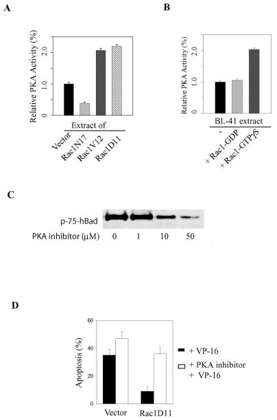
FIG. 6.
Rac1 stimulates PKA activity. (A) Equal amounts of extracts (2 μg) from healthy control BL-41 cells or from cell lines stably transfected with Rac1N17, Rac1V12, or Rac1D11 were subjected to the PepTag assay as described in the legend to Fig. 5. (B) Kinase activity was quantified by spectrophotometric analysis (absorbance at 570 nm) of the phosphorylated bands and is expressed relative to control (Vector alone) activity. (B) Purified Rac1 protein (2 μg) was preloaded with either GDP or GTPγS to inhibit or activate Rac1 activity, respectively. The Rac1 protein was then added to healthy BL-41 extracts and tested for an effect on PKA activity, which is expressed relative to the activity of active PKAC (10 ng). (C) Stable Rac1D11-expressing cells were treated for 60 min with an increasing concentration of a specific myristoylated PKA inhibitor. Whole-cell extracts were subjected to Western blotting to assess hBad Ser-75 phosphorylation. (D) Stable cell lines were pretreated with 10 μM PKA inhibitor prior to adding VP-16 (200 μg/ml). Apoptosis was allowed to proceed for 3 h. The percentage of apoptotic cells was assessed by fluorescence-activated cell sorting as described in the legend to Fig. 2. The data are representative of three independent assays.

