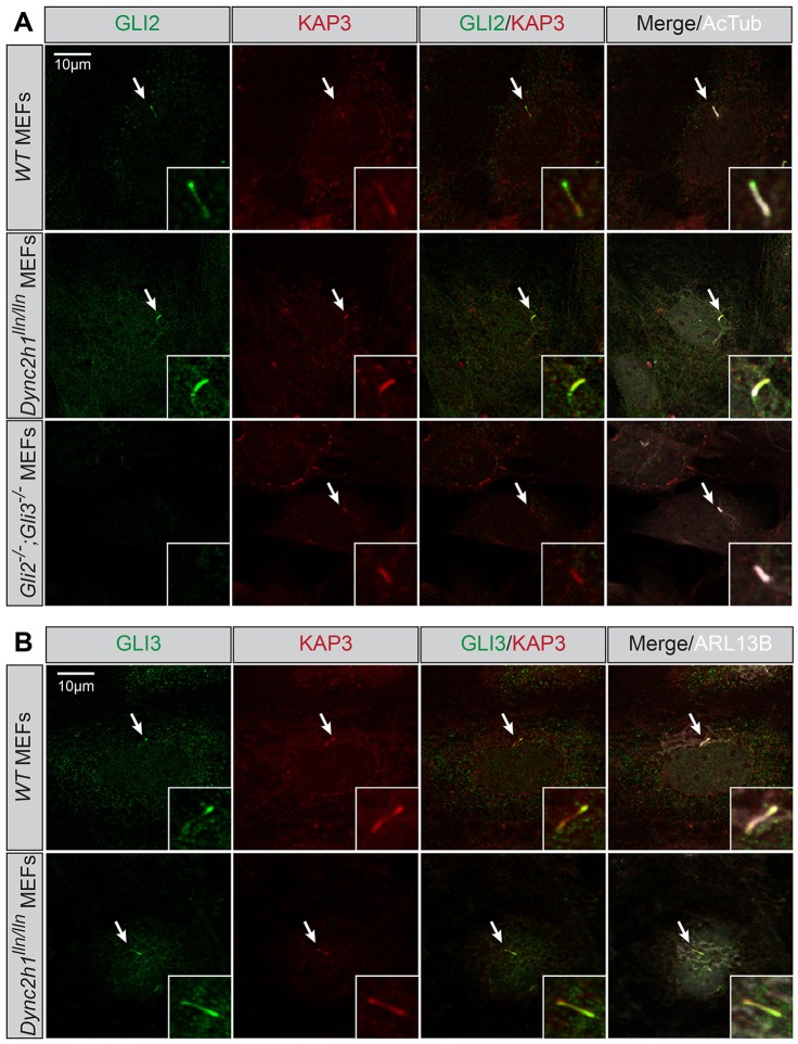
Fig. 1.
Endogenous KAP3 and GLI proteins localize to primary cilia. (A) Antibody detection of endogenous GLI2 (green) and KAP3 (red) in wild-type (WT, upper row), Dync2h1lln/lln (middle row) or Gli2−/−;Gli3−/− MEFs (lower row). Primary cilia are identified with antibodies against acetylated tubulin (AcTub; gray). (B) Antibody detection of endogenous GLI3 (green) and KAP3 (red) in wild-type (upper row) and Dync2h1lln/lln MEFs (lower row). Primary cilia are identified with antibodies against ARL13B (gray). Arrows denote localization of KAP3A and GLI2 or GLI3 in primary cilia. Insets show areas of interest at higher magnification.

