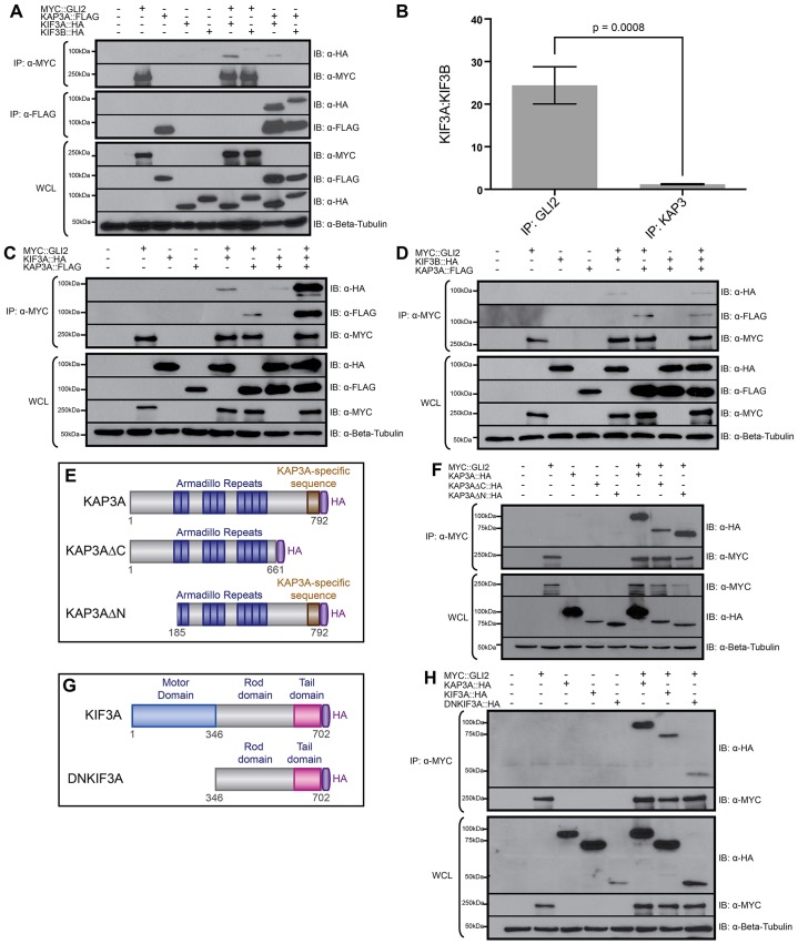
Fig. 3.
GLI2 selectively interacts with kinesin-2 motors and synergistically binds to the tail domain of KIF3A and the armadillo repeats of KAP3A. (A) Immunoprecipitation of MYC-tagged GLI2 (MYC::GLI2) or FLAG-tagged KAP3A (KAP3A::FLAG) from COS-7 cells expressing HA-tagged KIF3A (KIF3A::HA) or KIF3B (KIF3B::HA). (B) Quantification of the ratio of KIF3A:KIF3B that co-precipitates with either MYC::GLI2 or KAP3A::FLAG. Data show the mean±s.d. of the band densities from three separate experiments; a P-value of 0.0008 indicates a significant difference in the KIF3A:KIF3B ratio that co-precipitates with MYC::GLI2 compared with that co-precipitating with KAP3A::FLAG (Student's unpaired t-test). (C) Immunoprecipitation of MYC::GLI2 from COS-7 cells expressing KIF3A::HA and/or KAP3A::FLAG. (D) Immunoprecipitation of MYC::GLI2 from COS-7 cells expressing KIF3B::HA and/or KAP3A::FLAG. Immunoprecipitates (IP) and whole-cell lysates (WCL) were subjected to SDS-PAGE and western blot analysis (IB) using antibodies directed against MYC (α-MYC) and HA (α-HA). Antibody detection of β-tubulin (α-Beta-Tubulin) was used to confirm equal loading across lanes. (E) Schematic of full-length and truncated human KAP3A proteins. KAP3A contains highly conserved armadillo repeats (dark blue), a KAP3A-specific sequence (gold) and an HA-tag at the C-terminus (purple). (F) Immunoprecipitation of MYC::GLI2 from COS-7 cells expressing HA-tagged KAP3A (KAP3A::HA), KAP3AΔC (KAP3AΔC::HA) or KAP3AΔN (KAP3AΔN::HA). (G) Schematic of full-length and truncated human KIF3A proteins. KIF3A contains a highly conserved motor domain (blue), a rod domain (gray), a cargo-binding tail domain (pink) and an HA tag at the C-terminus (purple). (H) Immunoprecipitation of MYC::GLI2 from COS-7 cells expressing KAP3A::HA, KIF3A::HA or HA-tagged DNKIF3A (DNKIF3A::HA). Immunoprecipitates and whole-cell lysates were subjected to SDS-PAGE and western blot analysis using antibodies directed against MYC and HA. Antibody detection of β-tubulin was used to confirm equal loading across lanes. The molecular masses (in kDa) of protein standards are indicated at the left of each blot.

