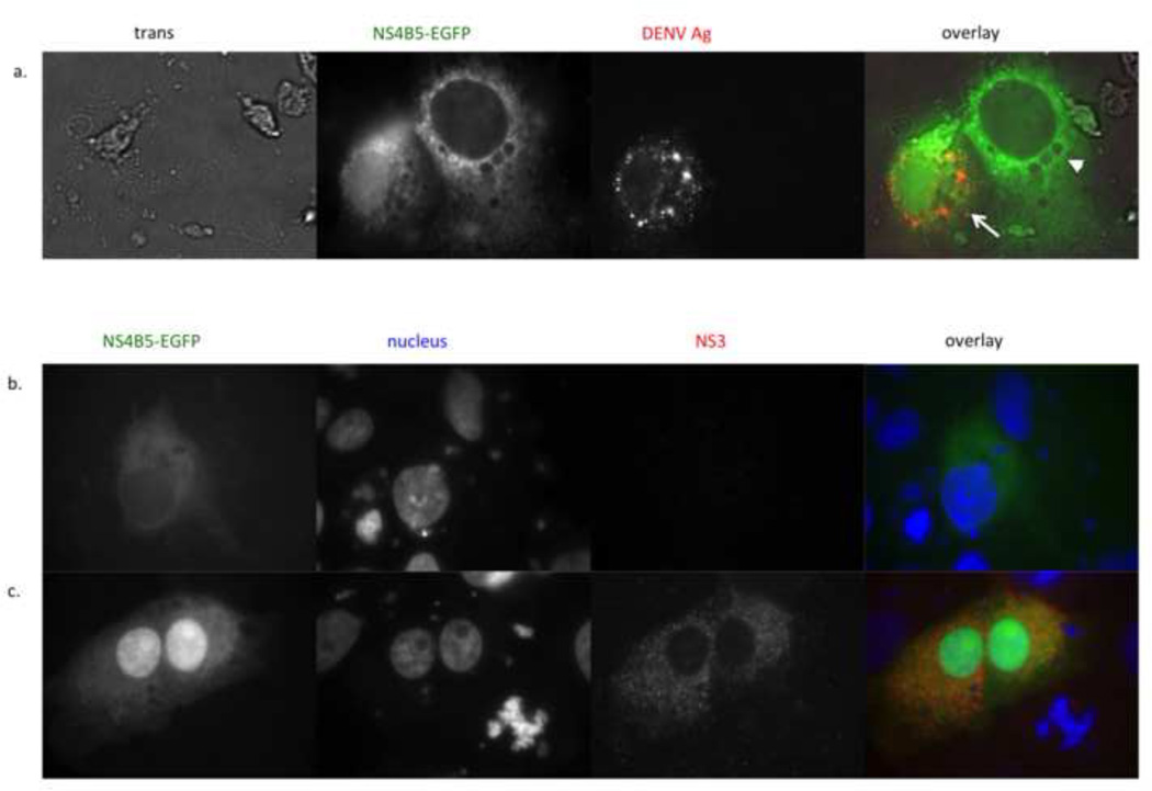
Figure 3.
Nuclear localization of GFP correlates with DENV antigen and co-expression of the NS2B3 protease. (a) Vero cells were transfected with p4B5-EGFP (green) and infected with DENV-2 16681 at an m.o.i of 1. 24 hours post-infection, cells were fixed, permeabilized and stained with antibody against DENV complex. The NS4B5-EGFP panel shows cytoplasmic expression of GFP (right, arrow neighboring cytoplasmic and nuclear expression of GFP (left, arrowhead) (magnification, ×100). DENV Ag panel shows DENV antigen staining in cells infected with DENV. Overlay of GFP and antigen staining (red) shows that nuclear localization of GFP correlates with DENV antigen staining (magnification, ×100). (b and c) Vero cells transfected with the p4B5-EGFP alone (b) or cotransfected with pNS2B3 (c) were analyzed for nuclear localization of GFP at 48hrs post-transfection (magnification, ×100). NS4B5-EGFP panel shows location of GFP within the cells (green). The nucleus panel shows the nucleus stained with NucBlue (blue, Life Technologies). The NS2B3 panel shows indirect antibody staining for NS3 (red). The overlay shows that nuclear GFP expression correlates with NS2B3 expression. Data are representative of at least six (a) and two (b) experiments.

