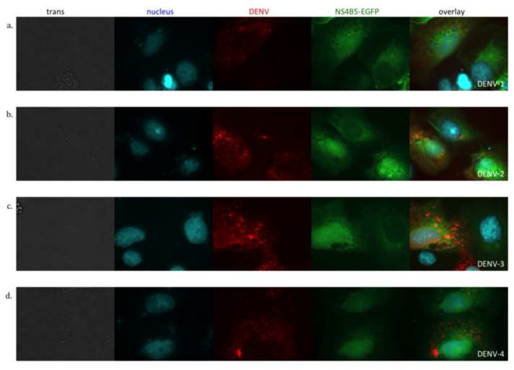
Figure 4.
All four DENV serotypes induce cleavage of p4B5-EGFP to localize GFP to the nucleus. Vero cells transfected with p4B5-EGFP (green) were infected with each of the four DENV serotypes at an MOI of 1. Cells were fixed, permeabilized and stained for DENV antigen (red) and nuclear DNA (cyan). Cells were analyzed at 24 hours post-infection by fluorescence microscopy. Each row is a representative image of DENV infected cells in bright field (trans) and fluorescence images showing nuclear stain (cyan), DENV antigen stain (red), 4B5-EGFP expression (green). The overlay is a composite of the nucleus, DENV and NS4B-EGFP images. (a) DENV-1 Hawaii, (b) DENV-2 C0112/96, (c) DENV-3 CH53489 and (d) DENV-4 814669. Data are representative of at least four experiments each.

