
Figure 1.
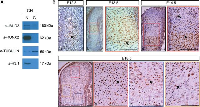
JMJD3 was highly expressed in prehypertrophic and hypertrophic chondrocytes. (A) Western blot analysis of JMJD3 in cytoplasmic (C) and nuclear (N) fractions of primary chondrocytes (CH) from E14.5 limbs. RUNX2 is a positive control. (B) Immunohistochemistry of JMJD3 at E12.5 (a), E13.5 (b, c), E14.5 (d, e), and E18.5 (f–i). WT humerus longitudinal sections counterstained with hematoxylin. Positive signal appears brown in the nuclear. The boxed regions in b, d, and f are magnified in c, e, g, h, and i with matched color rim, respectively. The black arrows indicate JMJD3-positive chondrocytes. Scale bar, 200 μm.