Figure 1.

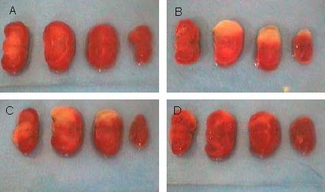
Morphology of brain coronal slices in mice. White part is the cerebral infarction lesion.
(A) In the sham-surgery group, brain tissue was red colored.
(B) In the model group, cerebral infarction was obviously visible.
(C) After ischemia, ketamine treatment failed to induce a significant change in infarct volume.
(D) After ischemia and reperfusion, ketamine treatment significantly decreased infarct volume.
