
Figure 1.
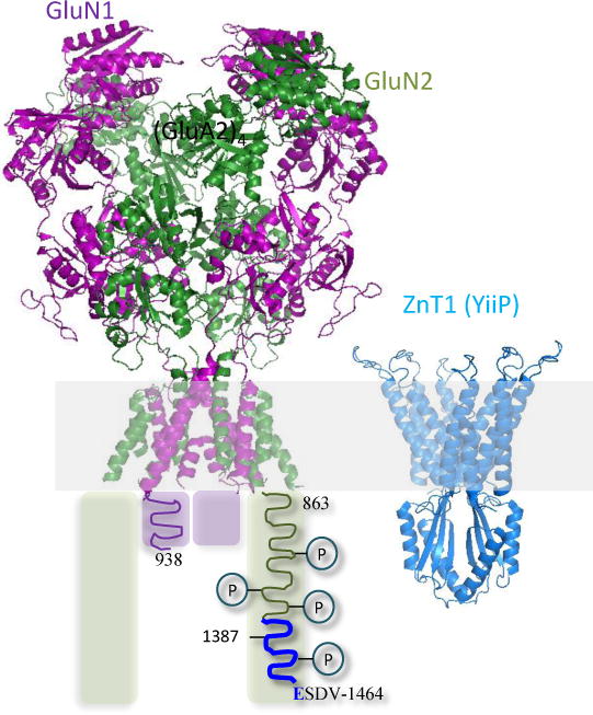
Structural models for an NMDA receptor and a zinc transporter. Left, Model of a tetrameric NMDA receptor combines the atomic arrangement for the ecto- and transmembrane domains of GluN1/GluN2B receptor (PDB 4PE5, Karakas and Furukawa, 2014) with a hypothetical cartoon of the cytoplasmic domain, which is presumed to be intrinsically disordered. Each GluN1 (purple) and GluN2A (green) subunit extends short (839 to 938 in GluN1) and long (863 to 1482, in GluN2A) intracellular tails; both include numerous phosphorylation sites; the sequence (1387–1461) required for interaction with ZnT1 is highlighted in blue. Right, Atomic model of a homo dimeric bacterial zinc transporter (YiiP, PDB: 3H90, Lu and Fu, 2007), which shares sequence homology with the mammalian zinc transporter ZnT1.