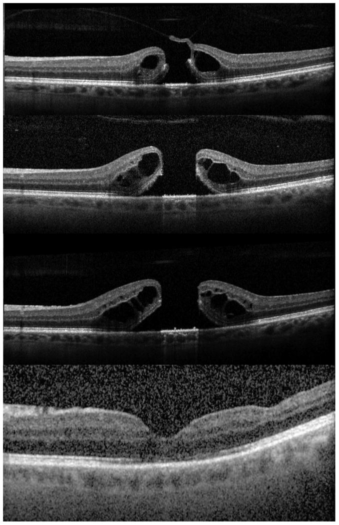
Figure 2.

Ocriplasmin failure A: Stage 2 macular hole prior to treatment. B: One week after ocriplasmin injection showing posterior vitreous detachment and enlargement of the macular hole. C: One month after ocriplasmin injection with further progression of intraretinal cystic changes and macular hole diameters. D: Seven months after surgical repair showing macular hole closure.
