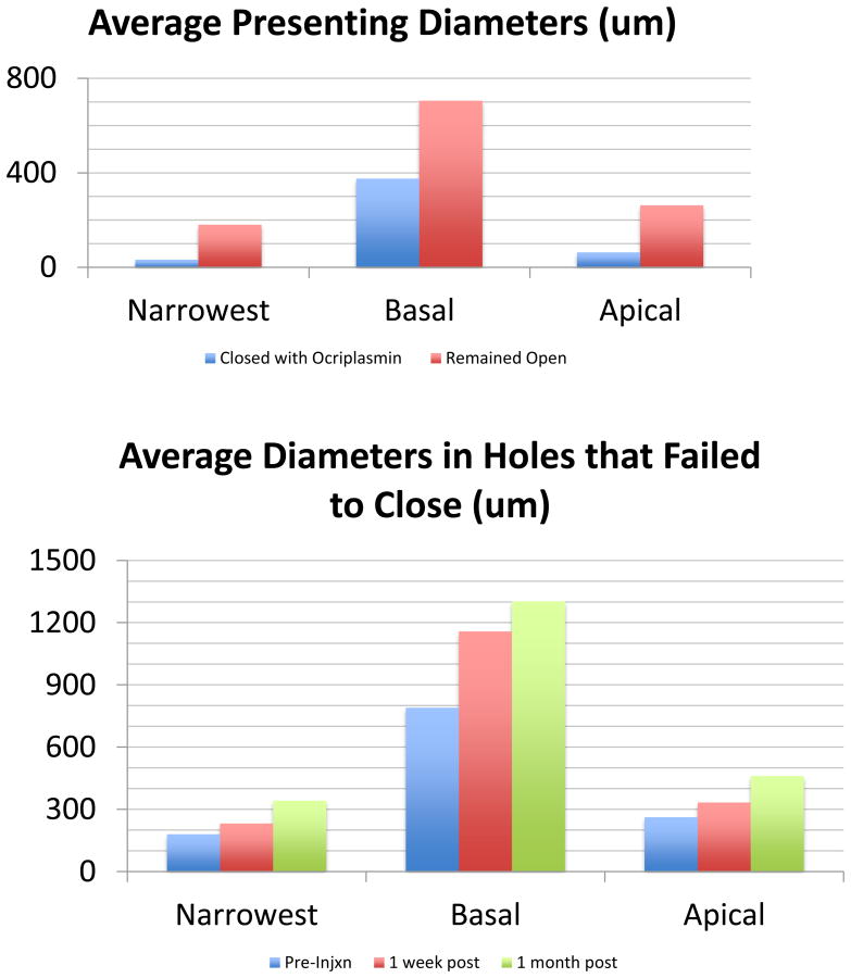
Figure 3.
OCT measurements A: The average diameters in the one hole that closed with ocriplasmin were smaller than those that failed to close. B: The average diameters of those holes that failed to close with ocriplasmin showed enlargement from pre-injection to 1 week post-injection to 1 month post-injection. These were statistically significant changes (*) in basal diameter at one week (p=0.002) and one month (p=0.006) and apical diameter at one month (p=0.016).

