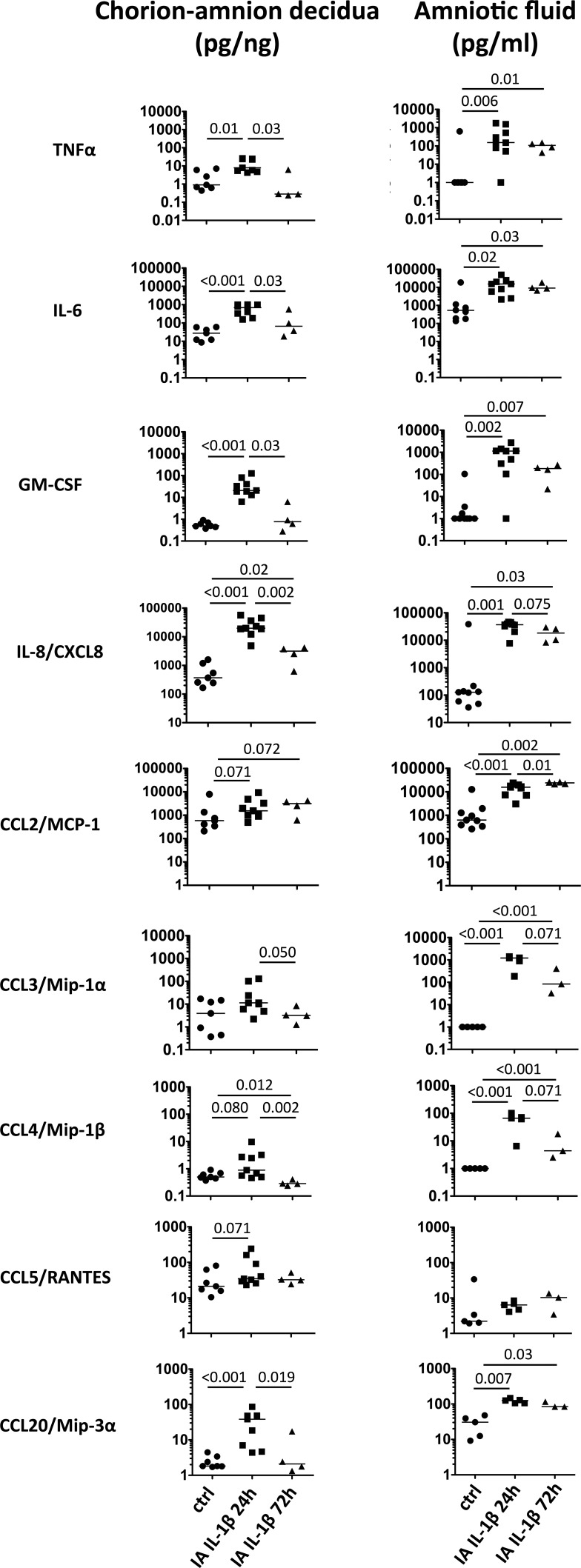
FIG. 1.
IA IL-1β increased inflammatory cytokines in chorioamnion-decidua and AF. Dot plots show cytokine levels in chorioamnion-decidua tissue homogenates and AF from controls (ctrl); IA IL-1β animals exposed for 24 h and 72 h. Horizontal bars correspond to medians, and P values are shown (n = 4–9 animals/group).

