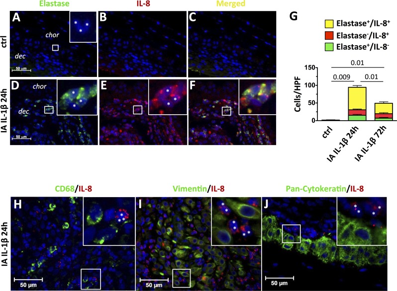
FIG. 5.
Neutrophils but not macrophages, decidua stromal cells, or fetal trophoblast cells produce IL-8 after IA IL-1β exposure. Chorioamnion-decidua sections from paraffin-embedded blocks were used for immunocolocalization studies and stained with neutrophil elastase (green) plus IL-8 (red) plus DAPI (blue). Representative sections are shown from control animals (A–C) and from animals exposed to IA IL-1β (D–F) at 24 h. In controls animals, cells with polymorphonuclear neutrophil morphology did not express neutrophil elastase (A) or IL-8 (B) or both (C). In contrast, IA IL-1β injection increased the proportion of polymorphonuclear cells (*inset) that expressed neutrophil elastase (D) or IL-8 (E). F) Merged images show colocalization of IL-8 and neutrophil elastase in the same cells. A, D, E, and F insets) Higher magnification pictures. G) Cell count/high powered field (HPF, ×40) of cytoplasmic neutrophil elastase+/IL-8−, elastase−/IL-8+, or neutrophil elastase+/IL-8+ (double-positive) cells in the chorion-decidua junction. P values for neutrophil elastase+/IL8+ neutrophils between the three groups of animals are shown. H–J) Representative sections from IA IL-1β 24-h animals of IL-8 colocalization and either macrophages (CD68 [green; H]), stromal cells (vimentin [green; I]), or trophoblast cells (pancytokeratin [green; J]). CD68+, vimentin+, or pancytokeratin+ cells do not colocalize with IL-8. In all panels, IL-8 expression is in red and DAPI is in blue. H, I, and J insets) Higher magnification picture, and white asterisks indicate IL-8+ polymorphonuclear cells. Original magnification ×4 (insets in A, D–F) and ×3 (insets in H–J).

