FIG. 6.
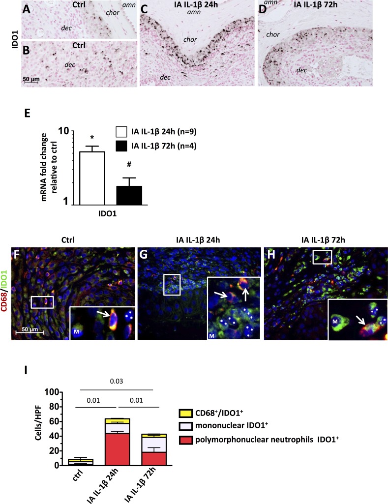
IA IL-1β exposure increased IDO1 production by neutrophils. A–D) Representative photomicrographs show immunohistology of IDO1-expressing cells in chorioamnion-decidua of control animals (A, B), those exposed to IA IL-1β after 24 h (C), and those exposed to IA IL-1β after 72 h. D) amn = amnion; chor = chorion; dec = decidua. E) Target mRNAs in chorioamnion-decidua were measured by quantitative RT-PCR and normalized to the expression of eukaryotic 18S rRNA. Values in experimental animals were expressed as fold change relative to that of a pooled average in the control animals (n = 5). *P < 0.05 compared to controls; #P < 0.05 compared to the group exposed to IA IL-1β at 24 h. IA IL-1β exposure increased IDO1. F–H) Representative photomicrographs from the merged frames from CD68 (green) plus IDO1 immunocolocalization. F) Higher magnification (insets ×40) views of samples from control animals show that only mononuclear cells (M) or CD68+ cells (white arrows) express IDO1, whereas IDO1+ cells are the predominant polymorphonuclear cells (nuclear lobes are shown by white asterisks) in IA IL-1β 24 h (G) and IA IL-1β 72 h (H) animals. I) Graph shows the cell count/HPF (×40) of IDO1+ cells in the choriodecidua junction: control animals show few CD68+IDO1+. In contrast, in IL-1β-treated animals, most polymorphonuclear neutrophils (nuclei indicated by white asterisks) are IDO1+. P values compare polymorphonuclear neutrophils IDO+ counts among the three groups of animals are shown. Original magnification ×3 (insets in F–H).

