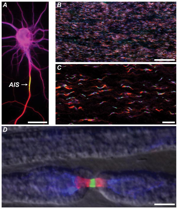
Figure 1. Examples of axonal patterning.
A. A cultured hippocampal neuron immunostained to mark the somatodendritic domain (magenta, MAP2), the axon initial segment (green, AnkG), and the axon (red, tubulin). Scale bar, 25 μm.
B. Low magnification view of optic nerve axons immunostained using antibodies against juxtaparanodal Kv1.2 (red), paranodal Caspr (green), nodal NF186 (Magenta). Scale bar, 50 μm.
C. High magnification view of optic nerve axons immunostained using antibodies against juxtaparanodal Kv1.2 (red), paranodal Caspr (green), nodal NF186 (magenta). Scale bar, 20 μm.
D. Triple immunostaining of PNS node of Ranvier immunostained for nodal Na+ channels (green), paranodal Caspr (red), and juxtaparanodal K+ channels (blue). A phase contrast image of the myelin sheath was merged with the fluorescence image to show the outline of the myelin sheath. Scale bar, 5 μm.

