
Figure 2.
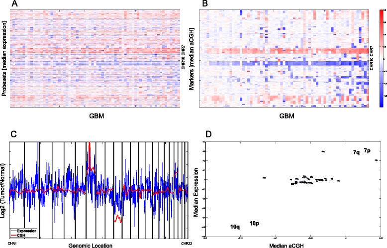
Correlation of gene expression and gene dosage across the GBM genome. Overall expression and aCGH in 64 GBM samples of the NCH-EORTC cohort are shown in fine resolution. (A) Gene expression is plotted relative to non-tumoral brain samples. Values were smoothened and interpolated. Every row is a probeset, and probesets are sorted by their genomic order. (B) aCGH data: Every row is a marker, and markers are sorted by their genomic order. In both A and B, the samples (columns) are sorted according to standard deviation in B. (C) For a single sample, the expression and aCGH values are shown. (D) For the same sample as in (C), the median of all markers on every chromosome arm is plotted against the median relative expression of all probesets on the same chromosomal arm. Only chromosome arms for which there are probesets and aCGH markers are shown.