
Figure 5.
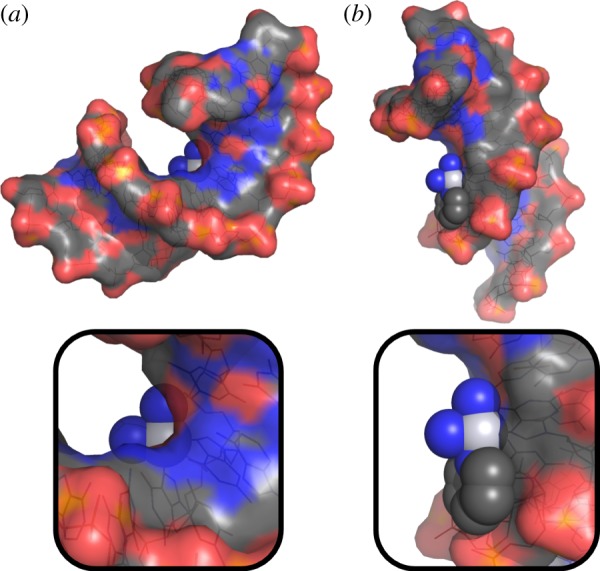
Top: surface representations of the cisplatin (a) and pyriplatin (b) adducts on duplex DNA with the atoms of the platinum lesion shown as spheres. The structures are oriented such that the platinum coordination plane lies in the plane of the page. Bottom: magnification of the platinum lesion illustrating the occlusion of the space above the platinum atom in the cisplatin lesion. (Online version in colour.)