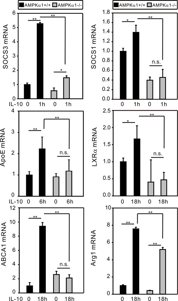
Figure 1.
AMPK mediates IL-10-induced gene expression in macrophages. BMDM generated from AMPKα1+/+ and AMPKα1−/− mice were treated with recombinant mouse (rm)-IL-10 (20 ng/ml) for the duration of 30 min, 1 h, 3 h, 6 h, and 18 h. Total cellular lysates were collected for real-time PCR analysis. The time point representing the peak level of expression for each gene is shown (representative result of two or more independent experiments). The mRNA expression of each gene was normalized to β-actin and compared to the AMPKα1+/+ untreated group. Data shown are the mean ± SD of triplicate determinations. Statistical significance between groups was calculated with an unpaired Student’s t test, with a value of p < 0.050 considered statistically significant. (**, p < 0.001. *, p < 0.050. n.s., p > 0.050).

