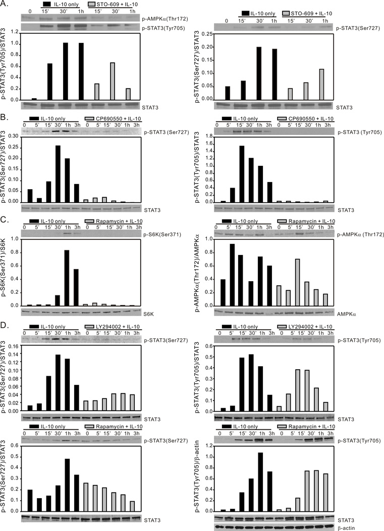
Figure 5.
IL-10 activation of STAT3 requires AMPK, JAK and mTORC1 activity. (A), BMDM generated from C57BL/6 mice were incubated with either media alone, or with the CaMKKβ inhibitor STO-609 (5 µM) for 1 h and then exposed to rm-IL-10 (20 ng/ml) for the time points indicated. Total cellular lysates were collected for Western blot assessment of AMPK-Thr172, STAT3-Tyr705 and STAT3-Ser727 phosphorylation. (B), BMDM generated from C57BL/6 mice were incubated with either media alone, or with JAK inhibitor CP-690550 (10 µM) for 1 h, then exposed to rm-IL-10 (20 ng/ml) for the time points indicated. Cell lysates were analyzed by Western blot for STAT3-Ser727 and STAT3-Tyr705 phosphorylation. (C), BMDM generated from C57BL/6 mice were incubated with either media alone, or with the mTORC1 inhibitor rapamycin (100 ng/ml) for 1 h and then exposed to rm-IL-10 (20 ng/ml) for the time points indicated. Total cellular lysates were collected for Western blot assessment of p70 S6K-Ser371 and AMPK-Thr172 phosphorylation. (D), BMDM generated from C57BL/6 mice were incubated with either media alone, with LY294002 (20 µM) for 1 h (top panels), or with the mTORC1 inhibitor rapamycin (100 ng/ml) for 1 h (bottom panels), then exposed to rm-IL-10 (20 ng/ml) for the time points indicated. Total cellular lysates were collected for Western blot assessment of STAT3-Ser727 and STAT3-Tyr705 phosphorylation. Protein phosphorylation levels were analyzed by densitometry and are displayed as a bar histogram. Results shown are representative of two to three independent experiments.

