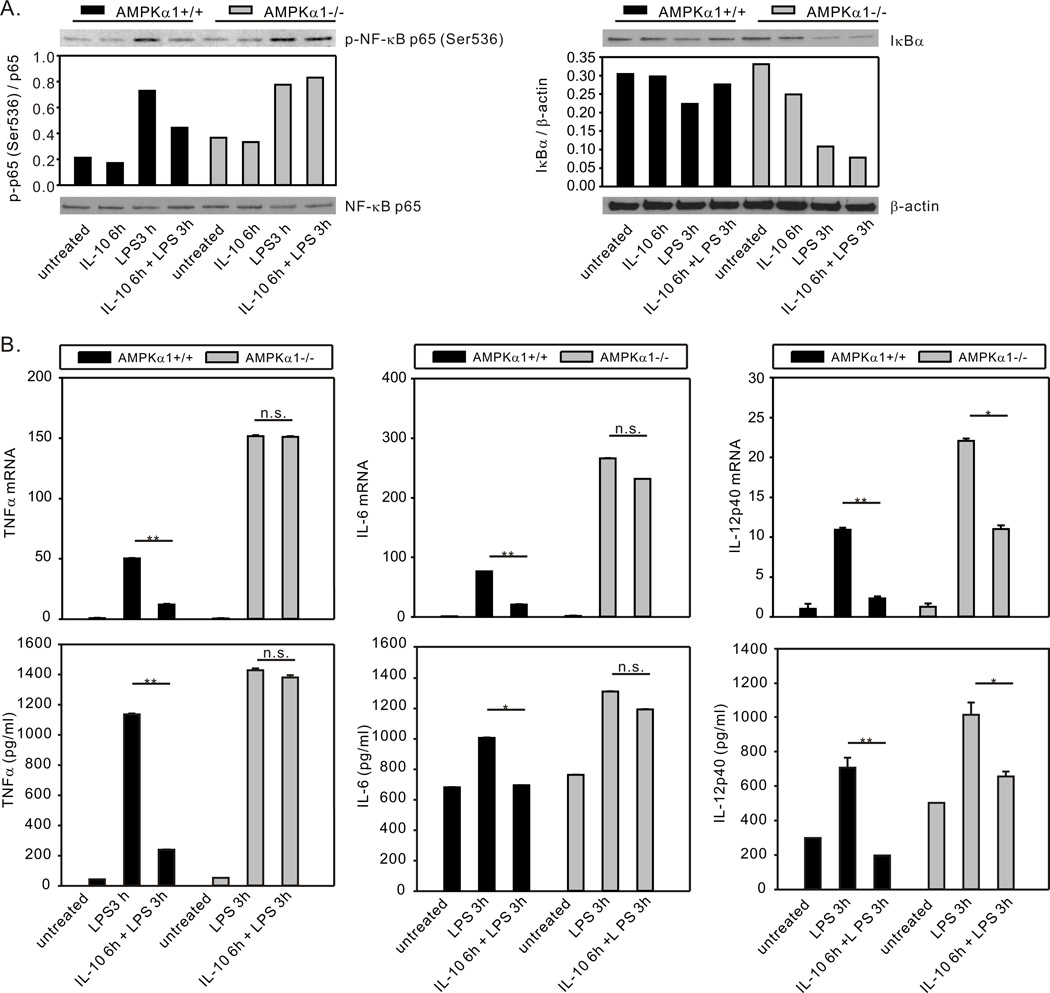
Figure 6.
AMPK contributes to IL-10-mediated suppression of LPS-induced NF-κB activation and proinflammatory cytokine production. (A), BMDM generated from AMPKα1+/+ and AMPKα1−/− mice were treated with rm-IL-10 (20 ng/ml) for 6h, then exposed to LPS (10 ng/ml) for 3 h. Total cellular lysates were collected for Western blot assessment of IκB degradation and NF-κB p65 phosphorylation. Protein expression or phosphorylation levels were analyzed by densitometry and are displayed as a bar histogram. (B), BMDM generated from AMPKα1+/+ and AMPKα1−/− mice were treated with rm-IL-10 (20 ng/ml) for 6h, then exposed to LPS (10 ng/ml) for 3 h. Total cellular lysates were collected for real-time PCR analysis of TNFα, IL-6 and IL-12p40 mRNA expression (top panels). The mRNA expression of each gene was normalized to β-actin and compared to the AMPKα1+/+ untreated group. Supernatants were collected for analysis by ELISA (bottom panels). The production level of each cytokine was compared to the AMPKα1+/+ untreated group. RT-PCR data shown are mean ± SD of triplicate determinations. ELISA data shown are mean ± SEM of triplicate determinations. Statistical significance between groups was calculated with an unpaired Student’s t test, with a value of p < 0.050 considered statistically significant. (**, p < 0.001. *, p < 0.050. n.s., p > 0.050). The data shown are representative of two or three independent experiments.

