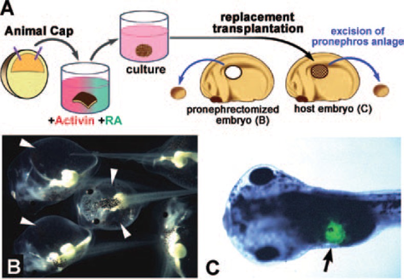
Fig. 6.
In vitro induction of pronephric tubules in animal caps. (A) The method for pronephros induction in animal caps and transplantation into the late neurula of Xenopus. Animal caps were treated with 10 ng/mL activin and 10−4 M of RA. The explant was implanted to replace the pronephros anlage. (B) Embryos from which the pronephric primordium has been excised are unable to eliminate water and clearly show edema (arrowheads). (C) An embryo transplanted with a pronephric explant shows normal development. Green fluorescent dye previously introduced into the explant indicates that it has been incorporated into the pronephric area (arrow).

