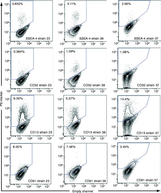
Figure 4.

Examples of FACS plots for cell surface markers that are heterogeneously expressed on tracheal basal cells. Primary human tracheal basal cells were stained with PE-conjugated antibodies for the indicated markers. FACS staining for strains 23 and 38 were done at the same time using the same control unstained cells and therefore, have the same gates. FACS staining for strain 37 was done at a different time and the gates for this strain are drawn relative to the unstained controls used at that time.
