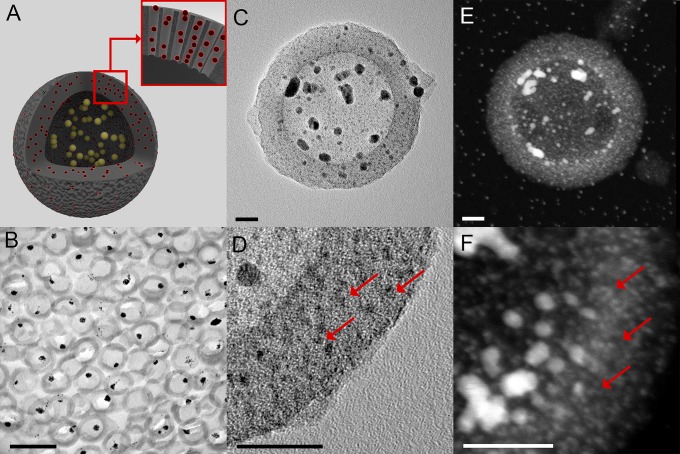
Fig. 1.
The QR’s morphology. (A) Schematic of a QR. QRs are composed of hollow mesoporous silica shells (HS, gray) hosting both AuQDs (red) inside their mesopores and gold nanoparticles AuNPs (yellow) inside their macrocavity. (B) Bright field TEM image of an ultrathin section of resin-embedded QRs, showing AuNPs within the mesoporous silica shells. (Scale bar, 0.2 µm.) (C) Bright field TEM image of a QR showing AuNPs within the cavity of the mesoporous silica shell. (Scale bar, 20 nm.) (D) Higher-magnification bright field TEM image of a QR silica shell showing the AuQDs (red arrows). (Scale bar, 20 nm.) (E) HAADF-STEM image of a QR highlighting the gold nanostructures within the particle. (Scale bar, 20 nm.) (F) Higher magnification HAADF-STEM image of a QR silica shell showing the AuQDs (red arrows). (Scale bar, 20 nm.)

