Fig. 3.
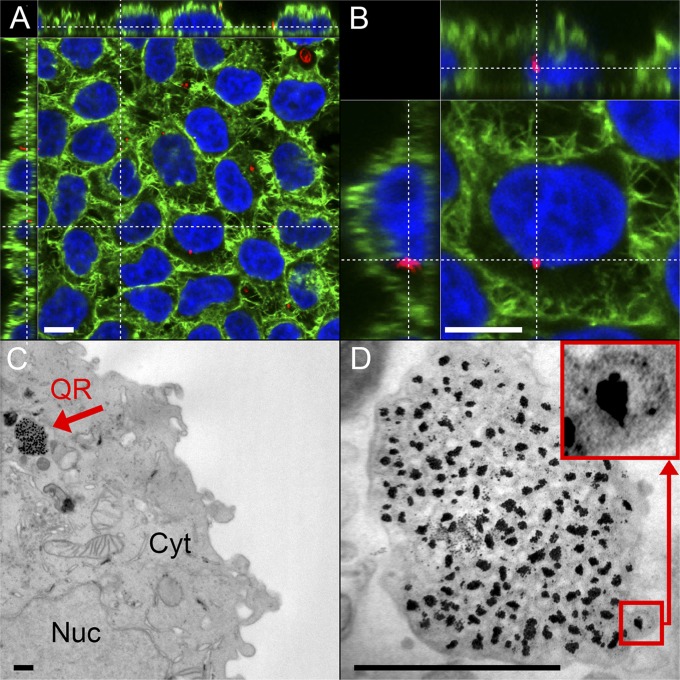
QR interaction with cells: internalization and NIR fluorescence in vitro. (A and B) Laser confocal scanning microscopy images of HeLa cells incubated for 1 d with QRs, which are imaged through their native fluorescence (red). Cells are stained for F-actin (AF488 phalloidin, green) and nucleus (DAPI, blue). QRs are localized by their native fluorescence within the cells and are found to accumulate in the cytoplasm and perinuclear region. (Scale bars, 10 µm.) Cell overview (A) and close-up of one HeLa cell (B), showing accumulation of QRs in the perinuclear region. An animated 3D reconstruction of the data set visualized in A and B is available as Movie S1. (C and D) Bright field TEMs of ultrathin sections of HeLa cells incubated with QRs for 1 d showing the internalization of the entire QR structure. (Scale bars, 500 nm.) (C) Cell overview (Cyt, cytoplasm; Nuc, nucleus). The red arrow points to the internalized QRs being trafficked within a vesicle. (D) Higher-magnification bright field TEM image of the QR agglomerate inside a vesicle. (Inset) An individual QR.

