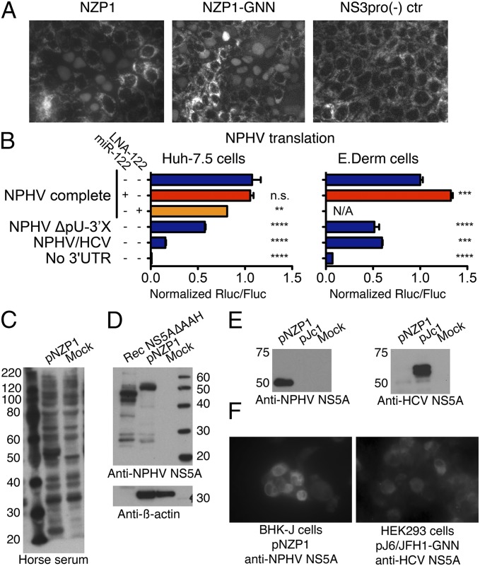
Fig. 3.
Translation of NPHV and detection of viral proteins. (A) Representative images of Clone8 cells 1 d posttransfection with RNA from NZP1, NZP1-GNN, or the protease-deficient control, NS3pro(−). (B) Translation from monocistronic translation reporters in Huh-7.5 or E.Derm cells. Relative Renilla (Rluc) to firefly (Fluc) luciferase values are shown after normalization for RNA amounts. Mean and SD are shown. Differences were evaluated by ANOVA. For P values, **P < 0.01, ***P < 0.001, and ****P < 0.0001. N/A, not applicable. (C) Western blot (WB) of lysates from T7-expressing HEK293 cells. Crude horse serum (lot 8211574) was used as a primary antibody. (D) WB of recombinant NS5A(ΔAAH) and the HEK293 lysates from B, using purified polyclonal NPHV NS5A8211574 antibody. The size difference for NS5A is due to absence of the amphipathic α-helix (AAH) in the recombinant protein. (E) NPHV and HCV NS5A antibodies do not cross-react. Lysates from T7-expressing HEK293 cells transfected with pNZP1 (NPHV) or pJc1 (HCV) were used for WB, using NPHV NS5A8211574 antibody or HCV NS5A9E10 antibody. (F) Immunostaining of T7-expressing HEK293 or BHK-J cells with and without NPHV (pNZP1) or HCV (pJ6/JFH1-GNN) detected with NPHV NS5A8211574 or HCV NS5A9E10 antibody.

