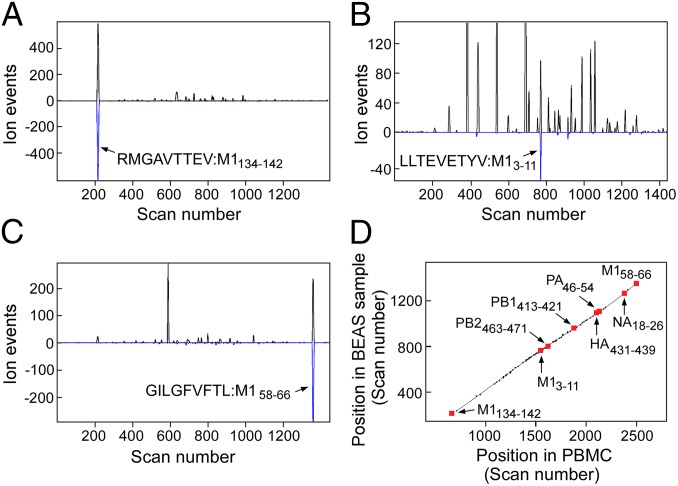
Fig. 1.
(A–C) Poisson detection plots of three M1 peptides extracted from affinity-purified HLA-A*02:01 complexes isolated from 5 million bronchial epithelial airway (BEAS-2B) cells infected with influenza strain A/Puerto Rico/8/1934. Black traces are extracted precursor ion chromatograms with the y scale being ion arrival events at the detector (counts) and the x scale, scan number (time). The blue trace is a Poisson chromatogram reflecting a likelihood of the peptide’s fragmentation pattern with units of scaled ion counts (SI Appendix, Outline of Poisson Segmented LC-DIAMS Method). Arrows mark the elution of the indicated peptide. (D) Elution mapping. Synthetic peptides of 31 targeted influenza epitopes were added to an extract of HLA-A2:01 bound peptides from 2.5 million A2+ peripheral blood mononuclear cells (PBMCs). Elution positions of major endogenous and synthetic peptides were recorded. When infected BEAS cells were analyzed, the elution positions of shared endogenous peptides and detected influenza epitopes were again recorded. Paired elution positions are plotted with the y axis as the scan position in the BEAS sample and the x axis, the position in PBMCs. Detected epitopes fall near the elution line.

