Fig. 1.
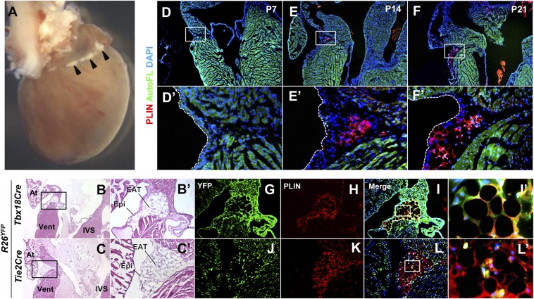
Tbx18Cre lineage-derived EAT is present in the AV groove of adult mice. (A) The dorsal surface of an adult (5 mo old) mouse heart. A small amount of fat (arrowheads) is visible in the AV groove. (B and C) Histological appearance of EAT. H&E-stained sections of adult mice show the presence of fat in the AV groove underneath the epicardium. The boxed regions are shown at higher magnification in B′ and C′. At, atrium; IVS, interventricular septum; Vent, ventricle. (D–F) Time course of appearance of AV groove fat. Staining with the adipocyte marker perilipin (PLIN) is absent until 2 wk after birth and increases with age. Autofluorescence (AutoFL) of the myocardium is included to delimit tissue borders. The boxed regions are shown at higher magnification in D′–F′. (G–L) Lineage origins of EAT adipocytes. The same sections are shown in the YFP (lineage marked) channel alone, the PLIN channel alone, or the merge of these and the DAPI channel. PLIN and YFP immunostaining colabel in Tbx18Cre lineage mapped tissue; small interstitial cells of unknown fate in the AV groove are labeled by Tie2Cre but these do not overlap PLIN+ adipocytes. The sections shown in G–L are adjacent to those used in B′ and C′. The boxed regions of I and L are shown at higher magnification in I′ and L′.

