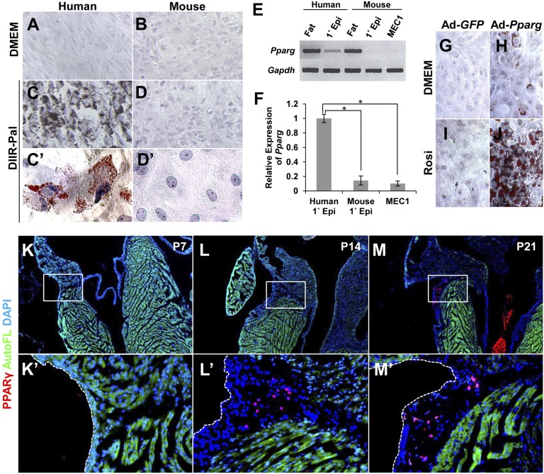
Fig. 2.
PPARγ controls epicardial adipogenesis. (A–D) In vitro adipogenic potential. Primary epicardial cells isolated from human and mouse embryonic ventricular tissue were cultured for 14 d in medium containing adipogenic inducers (dexamethasone, IBMX, insulin, rosiglitazone, palmitate) and stained with Oil Red O. (E) Pparg expression in epicardial cell cultures, analyzed by RT-PCR. Pparg was basally expressed in human cells, but was absent in mouse cells. Omental fat from adult human and adult mouse was used as positive controls. (F) Quantitation of normalized Pparg expression in human (n = 4 independent samples) and mouse (n = 3) primary epicardial cell cultures and MEC1 cell cultures (n = 3). The average human expression was set to 1.0. Statistically significant from human expression at *P = 0.0037 (mouse primary) and *P = 0.0012 (MEC1). (G–J) PPARγ transduction. PPARγ or GFP (used as a control) was transduced by adenovirus infection into primary mouse epicardial cells, and cells were then treated with rosiglitazone (Rosi). Lipid accumulation was visualized by Oil Red O staining. (K–M) PPARγ expression in vivo. PPARγ was detected in nuclei in the AV groove EAT of mouse heart 14 d after birth by immunostaining (red). Sections near those used for PLIN staining (Fig. 1 D–F) were used here for PPARγ immunostaining. The boxed regions are shown at higher magnification in K′–M′.

